Using the Catalyst methodology for innovating for impact, a multidisciplinary
and multi-sector team of faculty mentors will guide
participants through a structured process to consider real-world needs,
plausible solutions, and viable action plans to develop proposals that, if
successful, will lead to a clear and specific benefit
This Fall 2019 cohort will focus on Down Syndrome to produce research
and technology that give people with disabilities the possibility of
developing greater social and practical skills and improving their quality
of life in order to enhance their participation in the educational system,
in the workforce, and in community life.
Save the Date: Down syndrome research symposium Nov. 6th
Inaugural meeting will bring together local clinical and basic scientists studying Down Syndrome
Translational Research in Down Syndrome
1-6 pm November 6, 2019
Building 46 (43 Vassar St), MIT
We are excited to host our first DS research symposium!
The afternoon features talks by five Boston area researchers and clinicians, plus two keynote lectures, from Roger Reeves, Professor of Physiology at the Johns Hopkins University School of Medicine and principle investigator of the Down Syndrome Cognition Project, and Joaquin Espinosa, Professor of Pharmacology at University of Colorado Denver and Director of the Linda Crnic Institute for Down syndrome.
More details to follow!
ADSC researchers present at the T21RS conference
Come see our work at the international Down Syndrome meeting
The biennial Trisomy 21 Research Society International Conference is the premier scientific meeting for Down syndrome research, attracting basic and clinical scientists and practitioners from around the world. Four researchers from the ADSC will be presenting their work at this conference. If you are attending in Barcelona, please come visit us!
Friday, June 7, 12 pm
Talk, Dr. Hiruy Meharena (Tsai lab): “Consequences of trisomy 21 on the epigenome of different iPSC-derived cell types of the brain”
Friday, June 7, 4-6pm
Poster, John Replogle (Amon lab): “A CRISPR Screen to Identify Mutations that Relieve the Slow Growth of Trisomy 21 Cells “
Poster #: PO107
Saturday June 8, 8:30 am
Plenary Lecture, Dr. Li-Huei Tsai: “Alzheimer’s disease mechanisms and therapeutics”
(also, come meet her at the “Meet the Experts” session at 7 pm)
Sunday, June 9, 11 am
Short Talk, Becca Silberman (Amon lab): “Using Single-Cell Sequencing to Assess DNA Damage in Trisomy 21 Blood”
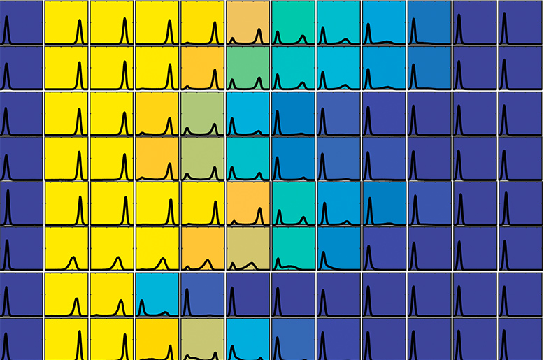
Colors represent variability of responses by cells with extra chromosomes
Aneuploidy is a condition in which cells contain an abnormal number of chromosomes, and is known to be the cause of many types of cancer and genetic disorders, including Down Syndrome. The condition is also the leading cause of miscarriage.
Disorders caused by aneuploidy are unusual in that the severity of their effects often varies widely from one individual to another.
For example, nearly 90 percent of fetuses with three copies of chromosome 21, the cause of Down Syndrome, will miscarry before birth. In other cases, people with the condition will live until they are over 60 years old.
Researchers have previously believed that this variation is the result of differences in the genetic makeup of those individuals with the condition.
But in a paper published today in the journal Cell, researchers at MIT reveal that aneuploidy alone can cause this significant variability in traits, in otherwise genetically identical cells.
The finding could have significant implications for cancer treatment, since it could explain why genetically identical cancer cells may respond differently to the same therapy.
An immediate impact
Aneuploidy originates during cell division, when the chromosomes do not separate properly or are not equally partitioned between the two daughter cells. This leads the cells, which in humans would normally have 46 chromosomes, to develop with either too many or too few chromosomes.
To study the effects of the condition, the researchers induced either chromosome loss or gain in genetically identical baker’s yeast cells. They chose baker’s yeast because the cells behave in a very similar way to human cells, according to Angelika Amon, the Kathleen and Curtis Marble Professor of Cancer Research, co-Director of the Alana Down Syndrome Center, and a member of the Koch Institute.
The induced changes had an immediate impact on the cells.
“We induced aneuploidy, and we found that the response was very variable from cell to cell,” Amon says. “Some cells slowed down their cycle completely, so that they could no longer divide, whereas others kept dividing quite normally and only experienced a small effect.”
The researchers carried out a systematic analysis, investigating the effect on the cells of gaining or losing a variety of different chromosomes. They found that in each case, even though individual cells had gained or lost the same chromosome, they behaved very differently from each other.
“So that really suggested that every single chromosome gained or lost had this effect, in that the responses (in each case) were quite variable,” Amon says.
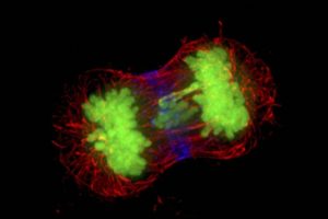
Microscopy image of dividing cells, with chromosomes in green. The chromosome in the middle is lagging, which can lead to incorrect chromosome number.
Beyond cell division
Microscopy image of dividing cancer cells, with chromosomes in green. The chromosome in the middle is lagging, which can lead to incorrect chromosome number.
The researchers also investigated the impact of aneuploidy on other biological pathways, such as transcription, the first stage of gene expression in which a segment of DNA is copied into RNA.
They found that here too, the effects of aneuploidy were varied across otherwise identical cells.
The cells’ response to environmental changes also varied considerably, suggesting that aneuploidy has an impact on the robustness of many, if not all, biological processes.
To ensure the response is not an effect that is unique to baker’s yeast cells, the researchers then studied the impact of aneuploidy on mice, and found the same levels of variability, Amon says.
“This suggests that the aneuploidy state itself could create variability, and that could provide an additional explanation of why diseases that are caused by aneuploidy are so variable,” Amon says.
Tumors, for example, are known to contain different populations of cells, some of which are quite different to each other in their genetic makeup. These genetic differences have often been blamed when chemotherapy or other treatments have been unsuccessful, as it was believed that the therapy may not have targeted all of the cells within the tumor.
“Unfortunately our paper suggests that tumors don’t even need to be heterogeneous genetically, the very fact that they have aneuploidy could lead to very variable outcomes, and that represents a significant challenge for cancer therapy,” Amon says.
Understanding the consequences of aneuploidy on cellular phenotypes is a fundamental question that has important implications for the treatment of several diseases, such as cancer and Down Syndrome, according to Giulia Rancati of the Institute of Medical Biology at the Agency for Science, Technology and Research (A*STAR) in Singapore, who was not involved in the research.
“This new exciting work adds an additional layer of understanding of how aneuploidy causes phenotypic variation, by revealing an unexpectedly high cell-to-cell variability between cells harboring the same aneuploidy karyotype,” Rancati says. “It would be interesting to test if this property of the aneuploid state might positively contribute to the evolution of cancer cells, which are known to develop drug resistance at high frequency.”
The researchers are now hoping to carry out further studies to investigate the origins of the variability, Amon says.
The results suggest that subtle changes in gene dosage across many genes, caused by the change in chromosome numbers, can promote alternate behaviors.
“We’re now trying to track down which the key genes are, and which the key pathways are,” she says. “Once we can understand what the key pathways are that cause this variability, we can start to think about targeting those pathways, to combat alternate outcomes in cancer treatment, for example.”
Helen Knight | MIT News correspondent
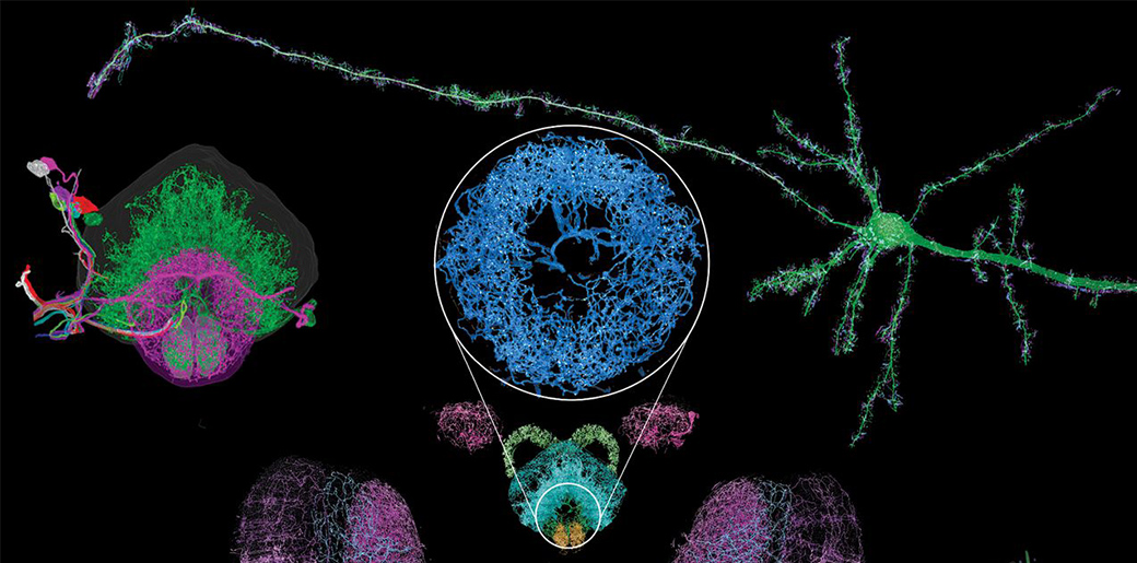
Mapping the brain at high resolution
New 3-D imaging technique can reveal, much more quickly than other methods, how neurons connect throughout the brain
Neural structures imaged using a new high-resolution, nanoscale imaging system.
Researchers have developed a new way to image the brain with unprecedented resolution and speed. Using this approach, they can locate individual neurons, trace connections between them, and visualize organelles inside neurons, over large volumes of brain tissue.
The new technology combines a method for expanding brain tissue, making it possible to image at higher resolution, with a rapid 3-D microscopy technique known as lattice light-sheet microscopy. In a paper appearing in Science Jan. 17, the researchers showed that they could use these techniques to image the entire fruit fly brain, as well as large sections of the mouse brain, much faster than has previously been possible. The team includes researchers from MIT, the University of California at Berkeley, the Howard Hughes Medical Institute, and Harvard Medical School/Boston Children’s Hospital.
This technique allows researchers to map large-scale circuits within the brain while also offering unique insight into individual neurons’ functions, says Edward Boyden, the Y. Eva Tan Professor in Neurotechnology, an associate professor of biological engineering and of brain and cognitive sciences at MIT, and a member of the Alana Down Syndrome Center.
“A lot of problems in biology are multiscale,” Boyden says. “Using lattice light-sheet microscopy, along with the expansion microscopy process, we can now image at large scale without losing sight of the nanoscale configuration of biomolecules.”
Boyden is one of the study’s senior authors, along with Eric Betzig, a senior fellow at the Janelia Research Campus and a professor of physics and molecular and cell biology at UC Berkeley. The paper’s lead authors are MIT postdoc Ruixuan Gao, former MIT postdoc Shoh Asano, and Harvard Medical School Assistant Professor Srigokul Upadhyayula.
Large-scale imaging
In 2015, Boyden’s lab developed a way to generate very high-resolution images of brain tissue using an ordinary light microscope. Their technique relies on expanding tissue before imaging it, allowing them to image the tissue at a resolution of about 60 nanometers. Previously, this kind of imaging could be achieved only with very expensive high-resolution microscopes, known as super-resolution microscopes.
In the new study, Boyden teamed up with Betzig and his colleagues at HHMI’s Janelia Research Campus to combine expansion microscopy with lattice light-sheet microscopy. This technology, which Betzig developed several years ago, has some key traits that make it ideal to pair with expansion microscopy: It can image large samples rapidly, and it induces much less photodamage than other fluorescent microscopy techniques.
“The marrying of the lattice light-sheet microscope with expansion microscopy is essential to achieve the sensitivity, resolution, and scalability of the imaging that we’re doing,” Gao says.
Imaging expanded tissue samples generates huge amounts of data — up to tens of terabytes per sample — so the researchers also had to devise highly parallelized computational image-processing techniques that could break down the data into smaller chunks, analyze it, and stitch it back together into a coherent whole.
In the Science paper, the researchers demonstrated the power of their new technique by imaging layers of neurons in the somatosensory cortex of mice, after expanding the tissue volume fourfold. They focused on a type of neuron known as pyramidal cells, one of the most common excitatory neurons found in the nervous system. To locate synapses, or connections, between these neurons, they labeled proteins found in the presynaptic and postsynaptic regions of the cells. This also allowed them to compare the density of synapses in different parts of the cortex.
Mouse neurons in yellow, with cyan and magenta markers for synapses, imaged with the new technique
MIT researchers have developed a method to perform large-scale, 3D imaging of brain tissue. Here, they image the entire fruit fly brain.
Using this technique, it is possible to analyze millions of synapses in just a few days.
“We counted clusters of postsynaptic markers across the cortex, and we saw differences in synaptic density in different layers of the cortex,” Gao says. “Using electron microscopy, this would have taken years to complete.”
The researchers also studied patterns of axon myelination in different neurons. Myelin is a fatty substance that insulates axons and whose disruption is a hallmark of multiple sclerosis. The researchers were able to compute the thickness of the myelin coating in different segments of axons, and they measured the gaps between stretches of myelin, which are important because they help conduct electrical signals. Previously, this kind of myelin tracing would have required months to years for human annotators to perform.
This technology can also be used to image tiny organelles inside neurons. In the new paper, the researchers identified mitochondria and lysosomes, and they also measured variations in the shapes of these organelles.
Circuit analysis
The researchers demonstrated that this technique could be used to analyze brain tissue from other organisms as well; they used it to image the entire brain of the fruit fly, which is the size of a poppy seed and contains about 100,000 neurons. In one set of experiments, they traced an olfactory circuit that extends across several brain regions, imaged all dopaminergic neurons, and counted all synapses across the brain. By comparing multiple animals, they also found differences in the numbers and arrangements of synaptic boutons within each animal’s olfactory circuit.
In future work, Boyden envisions that this technique could be used to trace circuits that control memory formation and recall, to study how sensory input leads to a specific behavior, or to analyze how emotions are coupled to decision-making.
“These are all questions at a scale that you can’t answer with classical technologies,” he says.
The system could also have applications beyond neuroscience, Boyden says. His lab is planning to work with other researchers to study how HIV evades the immune system, and the technology could also be adapted to study how cancer cells interact with surrounding cells, including immune cells.
The research was funded by K. Lisa Yang and Y. Eva Tan, John Doerr, the Open Philanthropy Project, the National Institutes of Health, the Howard Hughes Medical Institute, the HHMI-Simons Faculty Scholars Program, the U.S. Army Research Laboratory and Army Research Office, the US-Israel Binational Science Foundation, Biogen, and Ionis Pharmaceuticals.
Anne Trafton, MIT News Office
Brain Wave Stimulation May Improve Alzheimer’s Symptoms
A combination of light and sound can improve hallmarks of Alzheimer's in mice
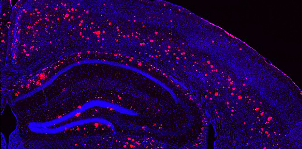
Microscope image of a light & sound treated mouse brain, cells labeled in blue, amyloid plaques in red.
By exposing mice to a unique combination of light and sound, MIT neuroscientists have shown that they can improve cognitive and memory impairments similar to those seen in Alzheimer’s patients. Individuals with Down syndrome have a high risk of developing Alzheimer’s Disease.
This noninvasive treatment, which works by inducing brain waves known as gamma oscillations, also greatly reduced the number of amyloid plaques found in the brains of these mice. Plaques were cleared in large swaths of the brain, including areas critical for cognitive functions such as learning and memory.
“When we combine visual and auditory stimulation for a week, we see the engagement of the prefrontal cortex and a very dramatic reduction of amyloid,” says Li-Huei Tsai, director of MIT’s Picower Institute for Learning and Memory and the Alana Down Syndrome Center, and the senior author of the study.
Further study will be needed, she says, to determine if this type of treatment will work in human patients. The researchers have already performed some preliminary safety tests of this type of stimulation in healthy human subjects.
MIT graduate student Anthony Martorell and Georgia Tech graduate student Abigail Paulson are the lead authors of the study, done in collaboration with Alana Investigator Ed Boyden’s lab, which appears in the March 14 issue of Cell.
Memory improvement
The brain’s neurons generate electrical signals that synchronize to form brain waves in several different frequency ranges. Previous studies have suggested that Alzheimer’s patients have impairments of their gamma-frequency oscillations, which range from 25 to 80 hertz (cycles per second) and are believed to contribute to brain functions such as attention, perception, and memory.
In 2016, Tsai and her colleagues first reported the beneficial effects of restoring gamma oscillations in the brains of mice that are genetically predisposed to develop Alzheimer’s symptoms. In that study, the researchers used light flickering at 40 hertz, delivered for one hour a day. They found that this treatment reduced levels of beta amyloid plaques and another Alzheimer’s-related pathogenic marker, phosphorylated tau protein. The treatment also stimulated the activity of debris-clearing immune cells known as microglia.
In that study, the improvements generated by flickering light were limited to the visual cortex. In their new study, the researchers set out to explore whether they could reach other brain regions, such as those needed for learning and memory, using sound stimuli. They found that exposure to one hour of 40-hertz tones per day, for seven days, dramatically reduced the amount of beta amyloid in the auditory cortex (which processes sound) as well as the hippocampus, a key memory site that is located near the auditory cortex.
“What we have demonstrated here is that we can use a totally different sensory modality to induce gamma oscillations in the brain. And secondly, this auditory-stimulation-induced gamma can reduce amyloid and Tau pathology in not just the sensory cortex but also in the hippocampus,” says Tsai, a founding member of MIT’s Aging Brain Initiative.
The researchers also tested the effect of auditory stimulation on the mice’s cognitive abilities. They found that after one week of treatment, the mice performed much better when navigating a maze requiring them to remember key landmarks. They were also better able to recognize objects they had previously encountered.
They also found that auditory treatment induced changes in not only microglia, but also the blood vessels, possibly facilitating the clearance of amyloid.
Dramatic effect
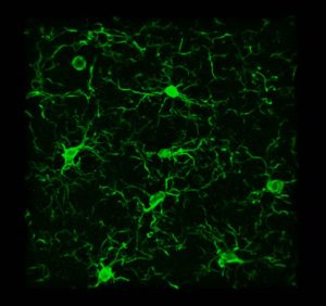
Brain cells called microglia, labeled green, change shape after light treatment
The researchers then decided to try combining the visual and auditory stimulation, and to their surprise, they found that this dual treatment had an even greater effect than either one alone. Amyloid plaques were reduced throughout a much greater portion of the brain, including the prefrontal cortex, where higher cognitive functions take place. The microglia response was also much stronger.
“These microglia just pile on top of one another around the plaques,” Tsai says. “It’s very dramatic.”
The researchers found that if they treated the mice for one week, then waited another week to perform the tests, many of the positive effects had faded, suggesting that the treatment would need to be given continually to maintain the benefits.
In an ongoing study, the researchers are now analyzing how gamma oscillations affect specific brain cell types, in hopes of discovering the molecular mechanisms behind the phenomena they have observed. Tsai says she also hopes to explore why the specific frequency they use, 40 hertz, has such a profound impact.
The combined visual and auditory treatment has already been tested in healthy volunteers, to assess its safety, and the researchers are now beginning to enroll patients with early-stage Alzheimer’s to study its possible effects on the disease.
“Though there are important differences among species, there is reason to be optimistic that these methods can provide useful interventions for humans,” says Nancy Kopell, a professor of mathematics and statistics at Boston University, who was not involved in the research. “This paper and related studies have the potential for huge clinical impact in Alzheimer’s disease and others involving brain inflammation.”
Anne Trafton, MIT News
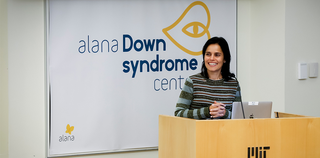
Alana Gift to MIT Launches Down Syndrome Research Center
Alana Down Syndrome Center will fund Down Syndrome biology research, technology program for disabilities
Alana Foundation President Ana Lucia Villela speaks at the Alana Down Syndrome Center launch event
As part of MIT’s continued mission to help build a better world, the Institute announced the creation of the Alana Down Syndrome Center, an innovative new research endeavor, technology development initiative, and fellowship program launched with a $28.6 million gift from Alana Foundation, a nonprofit organization started by Ana Lucia Villela of São Paulo, Brazil.
In addition to multidisciplinary research across neuroscience, biology, engineering, and computer science labs, the gift will fund a four-year program with MIT’s Deshpande Center for Technological Innovation called “Technology to Improve Ability,” in which creative minds around MIT will be encouraged and supported in designing and developing technologies that can improve life for people with different intellectual abilities or other challenges.
The Alana Down Syndrome Center, hosted out of MIT’s Picower Institute for Learning and Memory, will engage the expertise of scientists and engineers in a research effort to increase understanding of the biology and neuroscience of Down syndrome. The center will also provide new training and educational opportunities for early career scientists and students to become involved in Down syndrome research. Together, the center and technology program will work to accelerate the generation, development, and clinical testing of novel interventions and technologies to improve the quality of life for people with Down syndrome.
“At MIT, we value frontier research, particularly when it is aimed at making a better world,” says MIT President L. Rafael Reif. “The Alana Foundation’s inspiring gift will position MIT’s researchers to investigate new pathways to enhance and extend the lives of those with Down syndrome. We are grateful to the Foundation’s leadership — President Ana Lucia Villela and Co-President Marcos Nisti — for entrusting our community with this critical challenge.”
With a $1.7 million gift to MIT in 2015, Alana funded studies to create new laboratory models of Down syndrome and to improve understanding of the mechanisms of the disorder and potential therapies. In creating the new center, MIT and the Alana Foundation officials said they are building on that partnership to promote discovery and technology development aimed at helping people with different abilities gain greater social and practical skills to enhance their participation in the educational system, in the workforce, and in community life.
“We couldn’t be happier and more hopeful as to the size of the impact this Center can generate,” Villela said. “It’s an innovative approach that doesn’t focus on the disability but, instead, focuses on the barriers that can prevent people with Down Syndrome from thriving in life in their own way.”
Marcos Nisti, CEO & Vice-President of Alana, added, “This gift represents all the trust we have in MIT especially because the values our family hold are so aligned with MIT’s own values and its mission.”
Villela and Nisti have two daughters, one with Down syndrome. MIT Executive Vice President and Treasurer Israel Ruiz has had a personal connection to the Foundation.
“It is an extraordinary day,” Ruiz said. “It has been a pleasure getting to know Ana Lucia, Marcos and their family over the past few years. Their work to advance the needs of the Down Syndrome community is truly exemplary, and I look forward to future collaborations. Today, MIT celebrates their generosity in recognizing all abilities and working to provide opportunities to all.”
Down syndrome, also known as trisomy 21, is characterized by extra genetic material from some or all of chromosome 21 in many or all of an individual’s cells and occurs in one out of every 700 babies in the United States. Though the chromosomal hallmark of Down syndrome has been well known for decades, and advances in research, health care and social services have doubled lifespans over the past 25 years, significant challenges remain for individuals with different abilities and their families because the underlying neurobiology of the disorder is complex.
The center will be co-directed by Angelika Amon, the Kathleen and Curtis Marble Professor in Cancer Research, and Li-Huei Tsai, the Picower Professor of Neuroscience. Amon is an expert in understanding the health impacts of chromosomal instability and aneuploidy, the presence of an abnormal chromosome number, while Tsai is renowned for her work in the field of neurodegenerative disorders, including Alzheimer’s disease, which shares important underlying similarities with Down syndrome. In the first four years, the new center will employ cutting-edge techniques to study Down syndrome in the brain with two main focuses: systems and circuits as well as genes and cells.
With the support of the previous Alana Foundation gift, Hiruy Meharena, senior fellow in Tsai’s neuroscience lab, has already been deeply engaged in studying Down syndrome’s impact in the brain at the cellular and genomic level, examining key differences in gene expression in cultures of neurons and glia created from patient-derived induced pluripotent stem cells.
At the molecular and cellular level, Professor Manolis Kellis, director of MIT’s Computational Biology Group and a leader in big-data integration and analysis of genomic, epigenomic, and gene expression data will collaborate with Tsai for single-cell profiling of brain samples to understand the genes, molecular pathways, and cellular states that play causal roles in cognitive differences in Down syndrome.
At the systems and circuits level, Ed Boyden, the Y. Eva Tan Professor in Neurotechnology will lead efforts to conduct high-resolution 3D brain mapping and will collaborate with Tsai to examine the potential of using her emerging non-invasive, sensory-based therapy for Alzheimer’s in Down syndrome.
Amon’s lab will bring its deep expertise from their study of cancer to the new center. They have made important discoveries about how aneuploidy may undermine overall health, for instance by causing stresses within cells. It is their hope that identifying genetic alterations that suppress the stresses associated with trisomy 21 could lead to the development of therapeutics that improve cell function in individuals with Down syndrome.
To further support these research endeavors and to increase the long-term global pipeline of scientists trained in the study of Down syndrome, the Alana Down Syndrome Center will fund postdoctoral Alana Fellowships and graduate fellowships.
The Alana Center will also convene an annual symposium on Down syndrome research, the first of which is tentatively scheduled for this fall.
The Alana Foundation gift supports the MIT Campaign for a Better World, which was publicly launched in 2016 with a mission to advance MIT’s work in education, research, and innovation to address humanity’s urgent challenges. A joint statement guiding the gift’s purpose is available here.