Down Syndrome symposium presents bench-to-bedside research

First annual symposium brings together local Down Syndrome research community
Angelika Amon speaks at a podium
ADSC co-director Angelika Amon speaks about her work

At its first ever symposium Nov. 6, the Alana Down Syndrome Center demonstrated ways, from the scale of chromosomes to that of caregiving communities, that scientists and physicians in Massachusetts and around the country are working to help people with Down syndrome live their healthiest, fullest lives.

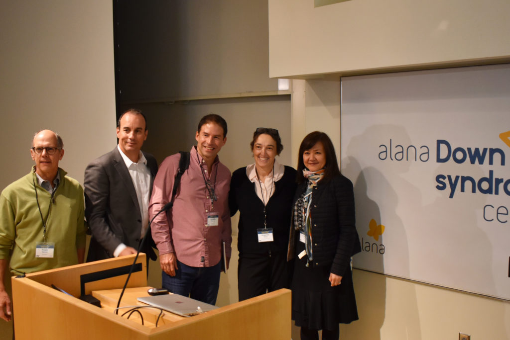
Founded at MIT in March 2019, the ADSC brings together neuroscience, biology, engineering and computer science research labs together with the Desphande Center for Technological Innovation to deepen knowledge about Down syndrome and to improve health, autonomy and inclusion of people with the genetic condition characterized by an extra copy of chromosome 21.

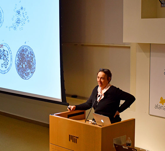
The symposium, “Translational Research in Down Syndrome,” brought together experts working across a spectrum of fundamental biology to clinical care.  In her opening remarks, ADSC co-director Li-Huei Tsai, Picower Professor of Neuroscience and director of The Picower Institute for Learning and Memory, said the event represented a chance for conversation and collaboration among researchers with the common goal of helping people with Down syndrome.

“Informed and inspired by their remarks we can all engage today in learning from each other,” she said. Tsai also thanked Ana Lucia Villela, whose Alana Foundation gift established the center and who had returned to MIT from Brazil to attend the symposium.

‘Aneuploidy’ advances

Throughout the afternoon, speakers shared some of their latest insights into how “aneuploidy,” having an atypical number of chromosomes, alters the biology of cells, the body and the brain.

One consequence appears to be that with an extra chromosome, cells make too many copies of the protein subunits that the chromosome encodes. Normally these subunits would become bound with partners encoded elsewhere into larger protein complexes, said ADSC Co-Director Angelika Amon, Kathleen and Curtis Marble Professor of Cancer Research in MIT’s biology department and the Koch Center for Integrative Cancer Research. But there aren’t as many of those partners, so the excess, unbound proteins become prone to clumping together, creating a major clean-up job for the cells that causes ”proteotoxic” stress. In Down syndrome, she said, that stress can hinder growth and proper function. Aneuploidy, she added, might also lead to a greater incidence of DNA damage.

Professors Reeves, Espinosa, Torres, Amon & Tsai pose for the camera
Professors Reeves, Espinosa, Torres, Amon & Tsai enjoy the meeting

Former Amon lab postdoc Eduardo Torres, who is now at the University of Massachusetts Medical School, said his lab has found that aneuploidy also disrupts the very shape and structure of the nucleus in a variety of cells, making them more sensitive to mechanical stress. The lab looked deeper to find the genetic and molecular pathway responsible and identified one related to the lipid composition of the nucleus. That insight allowed them to discover that administering certain drugs to cells with aneuploidy of chromosome 21 (or 13 or 18) can help shore up the nuclear structure and help cells grow.

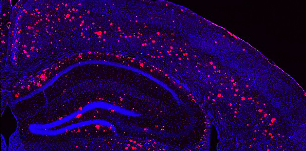
To gain more insight into how aneuploidy affects neurological development many scientists have begun using techniques to grow brain cells from stem cells derived from Down syndrome patients. They can manipulate these cultures in the lab so that the only genetic difference is the extra copy of chromosome 21. Jeanne Lawrence, also of UMass, said use of such advanced models will help her understand whether a technique her lab has developed to silence extra copies of a chromosome will be effective in cells such as those in the brain or blood. Her work shows promise for a potential gene therapy to mitigate the effects of the extra copy of chromosome 21.

Another vital model of Down syndrome is the mouse. In one of the day’s two keynote addresses, Roger Reeves of the McKusick-Nathans Institute for Genetic Medicine at Johns Hopkins University described what researchers have learned from the widely used T65dn mouse model, as well as what they hope to learn from a newly developed model, that uses human chromosome 21 genes to replicate chromosome duplication. He also described their studies of the developmental anomalies in Down syndrome model mouse brains, and have found that a crucial signaling pathway for development is less responsive in these mice. He reported the results of a screen to look for the specific contributors this pathway in DS, as they may be viable targets for drug development, and his lab has also identified some of these same genes to be involved in congenital heart defects.

Clinical care

In the symposium’s other keynote, Joaquin Espinosa of the Linda Crnic Institute for Down Syndrome at the University of Colorado, discussed how a fast-emerging raft of insights including discoveries about the immune system in Down syndrome has led to a new clinical trial. Fundamental research at the institute has found that patients with Down syndrome have an increased sensitivity to interferons, proteins emitted by immune cells as they fight infections. The research led scientists to test medicines to calm the immune system. He described their current work on a clinical trial that aims to investigate a drug, Xeljanz, already used for auto-immune disease, to see if the drug not only improves autoimmune skin diseases, but possibly a wider range of symptoms associated with Down syndrome.

Another clinical trial is getting underway at Boston Children’s Hospital, said Nicole Baumer, a researcher there who said there are real opportunities for interventions to improve cognition in Down syndrome patients, but who also cautioned that researchers must always consult patients and their caregivers about what they want from clinical trials and care, rather than assuming what’s best for them. After surveying to learn more about patient and family wishes, her group has designed a study in which they will try to predict the neurodevelopmental outcomes in babies with Down syndrome, and test whether behavioral therapy interventions designed for certain autism populations might also augment intellectual development in children with Down syndrome.

Dr. Brian Skotko presents a new program from his clinic

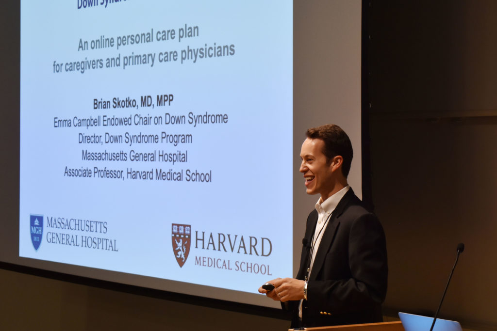
As researchers strive in the lab and clinic to make new discoveries and improve care, Brian Skotko of Massachusetts General Hospital and Harvard Medical School has also been considering how to ensure that state-of-the-art information reaches doctors and family caregivers everywhere it’s needed. Skotko noted that among approximately 212,000 people with Down syndrome in the United States, less than five percent have access to one of the 71 specialty clinics around the country like the one he directs at MGH. Instead, they typically depend on primary care physicians. That’s why he and a diverse team have spent the last two years developing an Internet-based platform, “Down Syndrome Clinic to You (DSC2U)” in which a physician or other caregiver can enter information about a patient and learn richly linked, expert-curated information and recommendations about medical care and wellness customized for the entered patient profile. The clinical team at MGH reviews the underlying database regularly to keep it up to date. With new data showing that the system positively influences care and has been valued by users, it’s ready for a wider launch next year, he said.

Taken together, the symposium talks illustrated many routes to potential progress, from the cell to the clinic.

Blending complementary expertise, Tsai and Kellis labs tackle brain diseases

Pair brings a team science approach to Down syndrome, Alzheimer's and other conditions
An illustration of a brain in profile overlaid with binary code

Li-Huei Tsai is a neuroscientist and Manolis Kellis is a computer scientist, so by working together, their research teams are able to ask questions about the big data of the brain that neither one could alone.

In their collaboration to help elucidate and mitigate Alzheimer’s disease and other neurological conditions, the labs of neuroscientist Li-Huei Tsai and computer scientist Manolis Kellis are two sides of the same coin on two sides of Vassar Street.

Bringing complementary skills to a shared mission as part of MIT’s Aging Brain Initiative and Alana Down Syndrome Center, the team seamlessly blends and advances some of the hottest and most powerful methods in science – statistical genetics, computational genomics, epigenomics, machine learning, single-cell profiling, “big data” integration, induced stem-cell reprogramming, mini-brain organoids, tissue engineering, and CRISPR-Cas9 genetic manipulation.This allows their teams to study genetic and molecular differences between healthy and diseased samples from multiple brain regions of humans and mice, integrate and analyze the resulting data to identify significant disease-driver genes and the cell types where they act, and engineer cells, tissues and mouse models to test their hypotheses and discover therapeutic interventions.

“Working together, we have the opportunity to garner big data from a large number of human subjects to elucidate the driver genes and pathways that are novel but key to the disease,” said Tsai, Picower Professor and director of the Picower Institute for Learning and Memory. “We can then test these genes/pathways in the induced pluripotent stem cells (iPSC) system coupled with CRISPR-Cas9 to manipulate the genome. We can induce the iPS cells into all major brain cell types, and dissect the contributions of each of these cell types to disease.”

It’s a joint research venture that’s as close, cutting-edge, and multidisciplinary as any at MIT, and fits squarely within the Schwarzman College of Computing’s emphasis on integrating artificial intelligence with the sciences. Kellis recalls it all getting started back in 2012 via the connection of postdocs, Elizabeth Gjoneska of the Tsai Lab and Andreas Pfenning from the Kellis Lab, who had met at a seminar on campus. With similarly overlapping interests in how gene regulation, and specifically epigenomic differences, affect the workings and health of the brain, they and other members of the two labs kindled dialogues that soon brought the professors together.

“The collaboration kind of happened organically,” said Kellis, professor of computer science and head of MIT’s Computational Biology Group. “We found kindred spirits – folks who thought similarly but were extremely complementary in their expertise.”

Within two years, the labs had jointly published two major papers. One in Nature, part of a sweeping set of reports on epigenomics that Kellis helped lead, showed that highly analogous sets of gene misregulation signals in the hippocampus of mice and humans each revealed a strong role for the brain’s immune cells and processes in allowing Alzheimer’s disease to progress. The other paper, in Cell, showed that in order to rapidly express genes critical for experience to affect synaptic connections, neurons naturally employ double-strand breaks of their DNA. The team hypothesized that failure to repair these breaks increases with age and may also contribute to neurodegeneration.

Each paper demonstrated the power of their combined approach. Since then, the collaboration has grown significantly to encompass about half a dozen projects. In 2016, for instance, they earned a National Institutes of Health grant to determine the significant epigenomic differences afoot in major brain cell types in Alzheimer’s disease.

In the last year, Kellis and Tsai received an influx of several new NIH grants and philanthropic gifts,  such as the one establishing the Alana Down Syndrome Center, enabling them to substantially expand their efforts in Alzheimer’s, tackle new disorders, bring in new collaborators, include new types of experiments, and expand their mechanistic studies. Their new directions include Schizophrenia, Bipolar Disorder, Psychosis in Alzheimer’s Disease, Frontotemporal Dementia, Lewy Body Dementia, and healthy aging.

Importantly, each experiment is designed together, Kellis says. Knowing that the team combines the capabilities of each lab, the team can be more ambitious.

“We think in a different way than any one lab would think by itself,” Kellis said. “For instance, I wouldn’t have the guts to ask many of these things that we are asking, if it wasn’t for our close collaboration with Li-Huei’s lab.”

In the Alana Center, they will apply their team science approach to modeling and analyzing Down syndrome, looking to identify and dissect the unique genetic and molecular signals that explain how the presence of an extra chromosome 21 affects the brain.

And with the new NIH grants, they will ask a litany of questions such as why many people with Alzheimer’s develop psychotic symptoms as well, what are the unique molecular signatures that distinguish Alzheimer’s and other dementias, and how do specific genetic variations in non-coding DNA elevate risk for a number of neurodegenerative and neuropsychiatric disorders.

“How privileged I feel to work with the world’s best computational team,” Tsai said. “This is only possible at MIT.”

Study helps explain varying outcomes for cancer, Down Syndrome

Differences in chromosome number may underlie variation among genetically identical individuals
Research Paper
A yellow to blue heat map
Colors represent variability of responses by cells with extra chromosomes

Aneuploidy is a condition in which cells contain an abnormal number of chromosomes, and is known to be the cause of many types of cancer and genetic disorders, including Down Syndrome. The condition is also the leading cause of miscarriage.

Disorders caused by aneuploidy are unusual in that the severity of their effects often varies widely from one individual to another.

For example, nearly 90 percent of fetuses with three copies of chromosome 21, the cause of Down Syndrome, will miscarry before birth. In other cases, people with the condition will live until they are over 60 years old.

Researchers have previously believed that this variation is the result of differences in the genetic makeup of those individuals with the condition.

But in a paper published today in the journal Cell, researchers at MIT reveal that aneuploidy alone can cause this significant variability in traits, in otherwise genetically identical cells.

The finding could have significant implications for cancer treatment, since it could explain why genetically identical cancer cells may respond differently to the same therapy.

An immediate impact

Aneuploidy originates during cell division, when the chromosomes do not separate properly or are not equally partitioned between the two daughter cells. This leads the cells, which in humans would normally have 46 chromosomes, to develop with either too many or too few chromosomes.

To study the effects of the condition, the researchers induced either chromosome loss or gain in genetically identical baker’s yeast cells. They chose baker’s yeast because the cells behave in a very similar way to human cells, according to Angelika Amon, the Kathleen and Curtis Marble Professor of Cancer Research, co-Director of the Alana Down Syndrome Center, and a member of the Koch Institute.

The induced changes had an immediate impact on the cells.

“We induced aneuploidy, and we found that the response was very variable from cell to cell,” Amon says. “Some cells slowed down their cycle completely, so that they could no longer divide, whereas others kept dividing quite normally and only experienced a small effect.”

The researchers carried out a systematic analysis, investigating the effect on the cells of gaining or losing a variety of different chromosomes. They found that in each case, even though individual cells had gained or lost the same chromosome, they behaved very differently from each other.

“So that really suggested that every single chromosome gained or lost had this effect, in that the responses (in each case) were quite variable,” Amon says.

Microscopy image of dividing cells, with chromosomes in green. The chromosome in the middle is lagging, which can lead to incorrect chromosome number.

Beyond cell division

2 dividing cells are labeled in blue & red, with green chromosomes being split between the two cells
Microscopy image of dividing cancer cells, with chromosomes in green. The chromosome in the middle is lagging, which can lead to incorrect chromosome number.

The researchers also investigated the impact of aneuploidy on other biological pathways, such as transcription, the first stage of gene expression in which a segment of DNA is copied into RNA.

They found that here too, the effects of aneuploidy were varied across otherwise identical cells.

The cells’ response to environmental changes also varied considerably, suggesting that aneuploidy has an impact on the robustness of many, if not all, biological processes.

To ensure the response is not an effect that is unique to baker’s yeast cells, the researchers then studied the impact of aneuploidy on mice, and found the same levels of variability, Amon says.

“This suggests that the aneuploidy state itself could create variability, and that could provide an additional explanation of why diseases that are caused by aneuploidy are so variable,” Amon says.

Tumors, for example, are known to contain different populations of cells, some of which are quite different to each other in their genetic makeup. These genetic differences have often been blamed when chemotherapy or other treatments have been unsuccessful, as it was believed that the therapy may not have targeted all of the cells within the tumor.

“Unfortunately our paper suggests that tumors don’t even need to be heterogeneous genetically, the very fact that they have aneuploidy could lead to very variable outcomes, and that represents a significant challenge for cancer therapy,” Amon says.

Understanding the consequences of aneuploidy on cellular phenotypes is a fundamental question that has important implications for the treatment of several diseases, such as cancer and Down Syndrome, according to Giulia Rancati of the Institute of Medical Biology at the Agency for Science, Technology and Research (A*STAR) in Singapore, who was not involved in the research.

“This new exciting work adds an additional layer of understanding of how aneuploidy causes phenotypic variation, by revealing an unexpectedly high cell-to-cell variability between cells harboring the same aneuploidy karyotype,” Rancati says. “It would be interesting to test if this property of the aneuploid state might positively contribute to the evolution of cancer cells, which are known to develop drug resistance at high frequency.”

The researchers are now hoping to carry out further studies to investigate the origins of the variability, Amon says.

The results suggest that subtle changes in gene dosage across many genes, caused by the change in chromosome numbers, can promote alternate behaviors.

“We’re now trying to track down which the key genes are, and which the key pathways are,” she says. “Once we can understand what the key pathways are that cause this variability, we can start to think about targeting those pathways, to combat alternate outcomes in cancer treatment, for example.”


Helen Knight | MIT News correspondent

Mapping the brain at high resolution

New 3-D imaging technique can reveal, much more quickly than other methods, how neurons connect throughout the brain
Research Paper
A neuron and several other colorful brain structures
Neural structures imaged using a new high-resolution, nanoscale imaging system.

Researchers have developed a new way to image the brain with unprecedented resolution and speed. Using this approach, they can locate individual neurons, trace connections between them, and visualize organelles inside neurons, over large volumes of brain tissue.

The new technology combines a method for expanding brain tissue, making it possible to image at higher resolution, with a rapid 3-D microscopy technique known as lattice light-sheet microscopy. In a paper appearing in Science Jan. 17, the researchers showed that they could use these techniques to image the entire fruit fly brain, as well as large sections of the mouse brain, much faster than has previously been possible. The team includes researchers from MIT, the University of California at Berkeley, the Howard Hughes Medical Institute, and Harvard Medical School/Boston Children’s Hospital.

This technique allows researchers to map large-scale circuits within the brain while also offering unique insight into individual neurons’ functions, says Edward Boyden, the Y. Eva Tan Professor in Neurotechnology, an associate professor of biological engineering and of brain and cognitive sciences at MIT, and a member of the Alana Down Syndrome Center.

“A lot of problems in biology are multiscale,” Boyden says. “Using lattice light-sheet microscopy, along with the expansion microscopy process, we can now image at large scale without losing sight of the nanoscale configuration of biomolecules.”

Boyden is one of the study’s senior authors, along with Eric Betzig, a senior fellow at the Janelia Research Campus and a professor of physics and molecular and cell biology at UC Berkeley. The paper’s lead authors are MIT postdoc Ruixuan Gao, former MIT postdoc Shoh Asano, and Harvard Medical School Assistant Professor Srigokul Upadhyayula.

Large-scale imaging

In 2015, Boyden’s lab developed a way to generate very high-resolution images of brain tissue using an ordinary light microscope. Their technique relies on expanding tissue before imaging it, allowing them to image the tissue at a resolution of about 60 nanometers. Previously, this kind of imaging could be achieved only with very expensive high-resolution microscopes, known as super-resolution microscopes.

In the new study, Boyden teamed up with Betzig and his colleagues at HHMI’s Janelia Research Campus to combine expansion microscopy with lattice light-sheet microscopy. This technology, which Betzig developed several years ago, has some key traits that make it ideal to pair with expansion microscopy: It can image large samples rapidly, and it induces much less photodamage than other fluorescent microscopy techniques.

“The marrying of the lattice light-sheet microscope with expansion microscopy is essential to achieve the sensitivity, resolution, and scalability of the imaging that we’re doing,” Gao says.

Imaging expanded tissue samples generates huge amounts of data — up to tens of terabytes per sample — so the researchers also had to devise highly parallelized computational image-processing techniques that could break down the data into smaller chunks, analyze it, and stitch it back together into a coherent whole.

In the Science paper, the researchers demonstrated the power of their new technique by imaging layers of neurons in the somatosensory cortex of mice, after expanding the tissue volume fourfold. They focused on a type of neuron known as pyramidal cells, one of the most common excitatory neurons found in the nervous system. To locate synapses, or connections, between these neurons, they labeled proteins found in the presynaptic and postsynaptic regions of the cells. This also allowed them to compare the density of synapses in different parts of the cortex.

Yellow cells with cyan and magenta dots
Mouse neurons in yellow, with cyan and magenta markers for synapses, imaged with the new technique

MIT researchers have developed a method to perform large-scale, 3D imaging of brain tissue. Here, they image the entire fruit fly brain.

Using this technique, it is possible to analyze millions of synapses in just a few days.

“We counted clusters of postsynaptic markers across the cortex, and we saw differences in synaptic density in different layers of the cortex,” Gao says. “Using electron microscopy, this would have taken years to complete.”

The researchers also studied patterns of axon myelination in different neurons. Myelin is a fatty substance that insulates axons and whose disruption is a hallmark of multiple sclerosis. The researchers were able to compute the thickness of the myelin coating in different segments of axons, and they measured the gaps between stretches of myelin, which are important because they help conduct electrical signals. Previously, this kind of myelin tracing would have required months to years for human annotators to perform.

This technology can also be used to image tiny organelles inside neurons. In the new paper, the researchers identified mitochondria and lysosomes, and they also measured variations in the shapes of these organelles.

Circuit analysis

The researchers demonstrated that this technique could be used to analyze brain tissue from other organisms as well; they used it to image the entire brain of the fruit fly, which is the size of a poppy seed and contains about 100,000 neurons. In one set of experiments, they traced an olfactory circuit that extends across several brain regions, imaged all dopaminergic neurons, and counted all synapses across the brain. By comparing multiple animals, they also found differences in the numbers and arrangements of synaptic boutons within each animal’s olfactory circuit.

In future work, Boyden envisions that this technique could be used to trace circuits that control memory formation and recall, to study how sensory input leads to a specific behavior, or to analyze how emotions are coupled to decision-making.

“These are all questions at a scale that you can’t answer with classical technologies,” he says.

The system could also have applications beyond neuroscience, Boyden says. His lab is planning to work with other researchers to study how HIV evades the immune system, and the technology could also be adapted to study how cancer cells interact with surrounding cells, including immune cells.

The research was funded by K. Lisa Yang and Y. Eva Tan, John Doerr, the Open Philanthropy Project, the National Institutes of Health, the Howard Hughes Medical Institute, the HHMI-Simons Faculty Scholars Program, the U.S. Army Research Laboratory and Army Research Office, the US-Israel Binational Science Foundation, Biogen, and Ionis Pharmaceuticals.

Anne Trafton, MIT News Office

Brain Wave Stimulation May Improve Alzheimer’s Symptoms

A combination of light and sound can improve hallmarks of Alzheimer's in mice
Research Paper
Microscope image of a light & sound treated mouse brain, cells labeled in blue, amyloid plaques in red.

By exposing mice to a unique combination of light and sound, MIT neuroscientists have shown that they can improve cognitive and memory impairments similar to those seen in Alzheimer’s patients. Individuals with Down syndrome have a high risk of developing Alzheimer’s Disease.

This noninvasive treatment, which works by inducing brain waves known as gamma oscillations, also greatly reduced the number of amyloid plaques found in the brains of these mice. Plaques were cleared in large swaths of the brain, including areas critical for cognitive functions such as learning and memory.

“When we combine visual and auditory stimulation for a week, we see the engagement of the prefrontal cortex and a very dramatic reduction of amyloid,” says Li-Huei Tsai, director of MIT’s Picower Institute for Learning and Memory and the Alana Down Syndrome Center, and the senior author of the study.

Further study will be needed, she says, to determine if this type of treatment will work in human patients. The researchers have already performed some preliminary safety tests of this type of stimulation in healthy human subjects.

MIT graduate student Anthony Martorell and Georgia Tech graduate student Abigail Paulson are the lead authors of the study, done in collaboration with Alana Investigator Ed Boyden’s lab, which appears in the March 14 issue of Cell.

Memory improvement

The brain’s neurons generate electrical signals that synchronize to form brain waves in several different frequency ranges. Previous studies have suggested that Alzheimer’s patients have impairments of their gamma-frequency oscillations, which range from 25 to 80 hertz (cycles per second) and are believed to contribute to brain functions such as attention, perception, and memory.

In 2016, Tsai and her colleagues first reported the beneficial effects of restoring gamma oscillations in the brains of mice that are genetically predisposed to develop Alzheimer’s symptoms. In that study, the researchers used light flickering at 40 hertz, delivered for one hour a day. They found that this treatment reduced levels of beta amyloid plaques and another Alzheimer’s-related pathogenic marker, phosphorylated tau protein. The treatment also stimulated the activity of debris-clearing immune cells known as microglia.

In that study, the improvements generated by flickering light were limited to the visual cortex. In their new study, the researchers set out to explore whether they could reach other brain regions, such as those needed for learning and memory, using sound stimuli. They found that exposure to one hour of 40-hertz tones per day, for seven days, dramatically reduced the amount of beta amyloid in the auditory cortex (which processes sound) as well as the hippocampus, a key memory site that is located near the auditory cortex.

“What we have demonstrated here is that we can use a totally different sensory modality to induce gamma oscillations in the brain. And secondly, this auditory-stimulation-induced gamma can reduce amyloid and Tau pathology in not just the sensory cortex but also in the hippocampus,” says Tsai, a founding member of MIT’s Aging Brain Initiative.

The researchers also tested the effect of auditory stimulation on the mice’s cognitive abilities. They found that after one week of treatment, the mice performed much better when navigating a maze requiring them to remember key landmarks. They were also better able to recognize objects they had previously encountered.

They also found that auditory treatment induced changes in not only microglia, but also the blood vessels, possibly facilitating the clearance of amyloid.

Dramatic effect

Brain cells called microglia, labeled green, change shape after light treatment

The researchers then decided to try combining the visual and auditory stimulation, and to their surprise, they found that this dual treatment had an even greater effect than either one alone. Amyloid plaques were reduced throughout a much greater portion of the brain, including the prefrontal cortex, where higher cognitive functions take place. The microglia response was also much stronger.

“These microglia just pile on top of one another around the plaques,” Tsai says. “It’s very dramatic.”

The researchers found that if they treated the mice for one week, then waited another week to perform the tests, many of the positive effects had faded, suggesting that the treatment would need to be given continually to maintain the benefits.

In an ongoing study, the researchers are now analyzing how gamma oscillations affect specific brain cell types, in hopes of discovering the molecular mechanisms behind the phenomena they have observed. Tsai says she also hopes to explore why the specific frequency they use, 40 hertz, has such a profound impact.

The combined visual and auditory treatment has already been tested in healthy volunteers, to assess its safety, and the researchers are now beginning to enroll patients with early-stage Alzheimer’s to study its possible effects on the disease.

“Though there are important differences among species, there is reason to be optimistic that these methods can provide useful interventions for humans,” says Nancy Kopell, a professor of mathematics and statistics at Boston University, who was not involved in the research. “This paper and related studies have the potential for huge clinical impact in Alzheimer’s disease and others involving brain inflammation.”

Anne Trafton, MIT News